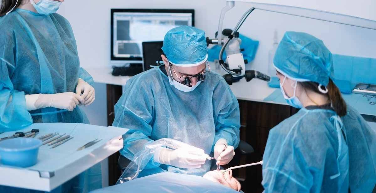
The face is the most visible part of the human body — and alterations to facial structure from cancer, trauma, or congenital deformity carry consequences that extend far beyond the physical. The appearance of the face shapes how people are perceived in social interactions, professional environments, and personal relationships. When cancer requires removal of facial bones, jaw, eye socket, or palate — or when trauma creates complex craniofacial injury — the reconstructive surgery that follows is not a cosmetic afterthought. It is a fundamental part of cancer treatment and quality of life restoration.
Craniofacial surgery in Ahmedabad sits at the intersection of oncological surgery, maxillofacial surgery, neurosurgery, and plastic reconstruction — one of the most technically complex fields in all of surgical medicine. At his practice, Dr. Vishal Choksi brings the subspecialty head and neck oncological surgical expertise that forms the cornerstone of craniofacial oncological reconstruction. As part of a dedicated cancer hospital in Ahmedabad team, and working alongside a specialist oncologist in Ahmedabad, Dr. Choksi ensures that patients undergoing craniofacial surgery receive both complete tumour removal and the best possible functional and aesthetic reconstruction.
What Is Craniofacial Surgery?
Craniofacial surgery encompasses surgical procedures involving the bones and soft tissues of the cranium (skull) and face — including the orbits (eye sockets), jaws, palate, nasal bones, zygomatic arches, frontal bone, and the skull base. In oncological practice, craniofacial surgery is predominantly reconstructive — restoring structure and function after cancer resection — though it also addresses traumatic craniofacial injuries and, in the paediatric population, congenital craniofacial deformities (craniosynostosis, cleft lip and palate, orbital hypertelorism).
Oncological Indications for Craniofacial Surgery in Ahmedabad
Oral and Maxillary Cancer Requiring Jaw Reconstruction
Oral cavity cancers — squamous cell carcinoma of the tongue, floor of mouth, buccal mucosa, gingiva, hard palate, and retromolar trigone — may invade the mandible (lower jaw) or maxilla (upper jaw). When bone invasion is confirmed or strongly suspected on imaging, resection must include the involved bone segment.
Mandibular resection (mandibulectomy):
- Marginal mandibulectomy: The outer cortex of the mandible is removed while preserving the inferior border and continuity — for periosteal invasion without cortical penetration
- Segmental mandibulectomy: A segment of the mandible is removed, interrupting jaw continuity — for cortical or medullary bone invasion
After segmental mandibulectomy, jaw reconstruction restores both the bite (dental occlusion) and facial contour. The gold standard is the fibula free flap — a segment of fibula bone (with its overlying skin paddle and periosteum) transferred on its peroneal vascular pedicle via microvascular anastomosis to the neck vessels. The fibula provides excellent bone stock, can be osteotomised to match mandibular curvature, and can be pre-operatively planned using virtual surgical planning (VSP) and custom cutting guides.
For oral and jaw cancer treatment: Mouth Cancer in Ahmedabad — Dr. Vishal Choksi
Total or Subtotal Maxillectomy for Upper Jaw Cancer
Cancers of the hard palate, maxillary gingiva, or maxillary antrum (upper sinus) may require partial or total maxillectomy — removal of part or all of the upper jaw. This creates a communication between the oral and nasal cavities (oronasal fistula) and a significant facial contour defect.
Reconstruction options include:
- Prosthetic obturator — a removable dental plate that obliterates the defect; simpler but with functional limitations
- Free flap reconstruction — anterolateral thigh or rectus abdominis flap with split-thickness skin graft lining; more complex but provides permanent reconstruction with better functional outcomes
- Fibula osteocutaneous free flap — for cases where dental rehabilitation with osseointegrated implants is planned
Orbital Exenteration and Orbital Reconstruction
Orbital exenteration — removal of the orbital contents including the eye — is required when cancer invades the orbit from adjacent structures (sinonasal cancer, skin cancer, lacrimal gland tumour). Reconstruction of the orbital defect is a major craniofacial undertaking, involving:
- Coverage of the orbital cavity with a temporalis muscle transposition or free flap
- Orbital prosthesis fitting (post-healing)
- Osseointegrated implants for prosthesis retention
Anterior Skull Base Resection and Reconstruction
For sinonasal cancers with cribriform plate invasion, the resection specimen includes the anterior skull base — creating a dural defect communicating the cranial cavity with the nasal cavity. Reconstruction of this defect to prevent cerebrospinal fluid leak and ascending meningitis is a critical part of the craniofacial operation:
- Pericranial flap — a scalp-based flap used to seal the skull base defect from above
- Free flap with dural reconstruction — for larger defects where a pericranial flap is insufficient
- Titanium mesh — for bony skull base reconstruction when required
For skull base surgery service: Skull Base Cancer Surgeon in Ahmedabad
The Role of Microvascular Free-Flap Reconstruction in Craniofacial Surgery
The most significant advance in craniofacial reconstruction oncology over the past three decades is the routine availability of microvascular free-flap surgery — the transfer of a block of tissue (skin, muscle, bone, or a combination) from a distant donor site to the face and jaw, connected to the recipient blood supply in the neck by microsurgical arterial and venous anastomoses.
Commonly used free flaps in head and neck and craniofacial reconstruction:
Fibula osteocutaneous free flap: The workhorse of mandible and maxilla reconstruction. The fibula provides excellent bone length, strength, and adaptability. The skin paddle monitors flap viability post-operatively. Donor site is well-tolerated with appropriate physiotherapy.
Anterolateral thigh (ALT) free flap: A versatile soft tissue flap from the lateral thigh. Can be harvested as a thin fasciocutaneous flap or as a thicker musculocutaneous flap. Used for pharyngeal reconstruction, palate reconstruction, and oral cavity lining.
Radial forearm free flap: A thin, pliable fasciocutaneous flap from the forearm. Ideal for tongue reconstruction, floor of mouth reconstruction, and cheek lining — where thin tissue with good sensory recovery is preferred.
Pectoralis major myocutaneous pedicled flap: Not a free flap — the blood supply is maintained from the thoracoacromial pedicle. Less reliable for complex craniofacial reconstruction but useful as a backup and for neck coverage.
Recovery and Rehabilitation After Craniofacial Surgery
Craniofacial reconstruction is among the most physiologically demanding operations in head and neck surgery. Patients require:
- ICU monitoring in the immediate post-operative period (typically 24–48 hours) for flap viability monitoring and airway management
- Regular flap observation — hourly for the first 48 hours; changes in skin paddle colour, temperature, or Doppler signal prompt urgent return to theatre for salvage
- Physiotherapy — jaw opening exercises after mandibular reconstruction to prevent trismus
- Speech and swallowing therapy — particularly after tongue, palate, or pharyngeal reconstruction
- Nutritional support — nasogastric or jejunostomy feeding until oral intake is re-established
- Dental rehabilitation — osseointegrated implant placement (6–12 months after reconstruction) for patients with fibula mandible reconstruction
For life quality after major head and neck cancer surgery: Life After Head and Neck Cancer Treatment in Ahmedabad
Conclusion: Craniofacial Surgery in Ahmedabad — Where Oncological Clearance Meets Reconstructive Excellence
Craniofacial surgery in Ahmedabad is only as good as the surgeon performing it — and for oncological craniofacial surgery, the best outcomes are produced by a surgeon who combines the oncological precision to achieve tumour-free margins with the reconstructive expertise to restore function and form. At Dr. Vishal Choksi’s oncological surgery practice — working within a cancer hospital in Ahmedabad infrastructure and alongside a multidisciplinary oncologist in Ahmedabad team — patients receive both components under unified expert care.
Book Your Craniofacial Surgery Consultation
Major craniofacial reconstruction for cancer requires subspecialty expertise — and adequate time for surgical planning before operation.


Leave a comment